Techniques
- Biological samples imaging using transmissive electron microscopy (TEM).
- Cryo EM cryogenic imaging using transmissive electron microscopy.
- Energy dispersive X-ray spectroscopy – element sample analysis.
- Ultrastructure samples creation in both room temperature and cryogenic conditions.
- Immunogold labeling.
Laboratory equipment
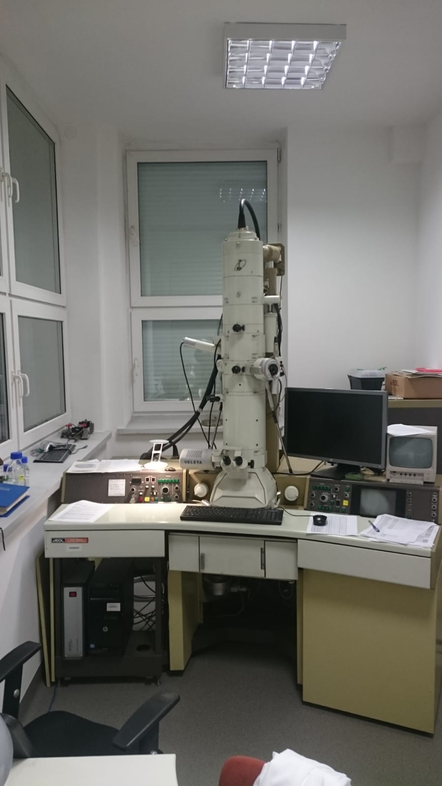
- JEOL 1200 EX microscope,
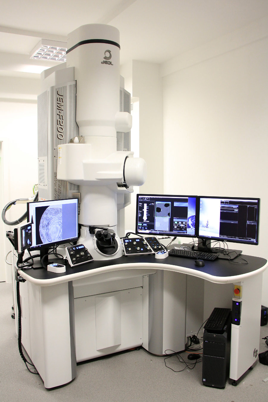
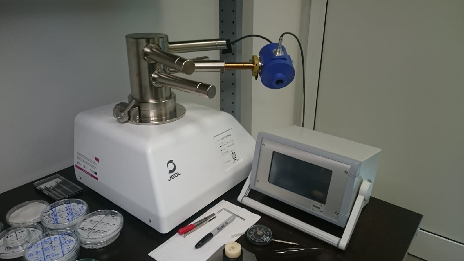
- JEOL JEM F-200 microscope,
- Cold field emission with maximum accelerating voltage of 200 kV,
- CR pole piece, with maximum available contrast and low contamination parameter, dedicated for low temperature and general biological imaging,
- EDS JEOL CENTURIO DRY SD30GV detector
- STEM microscope mode with Bright Field (BF) detectors and High-Angle Annular Dark-Field (HAADF),
- CMOS TVIPS TemCam-XF416 camera with 16 MPix resolution and 5 x 63.5 mm2 area,
- High tilt handle for room temperature tomography,
- FISCHIONE 2550 Cryo-Transfer low temperature handle with tomography capabilities.
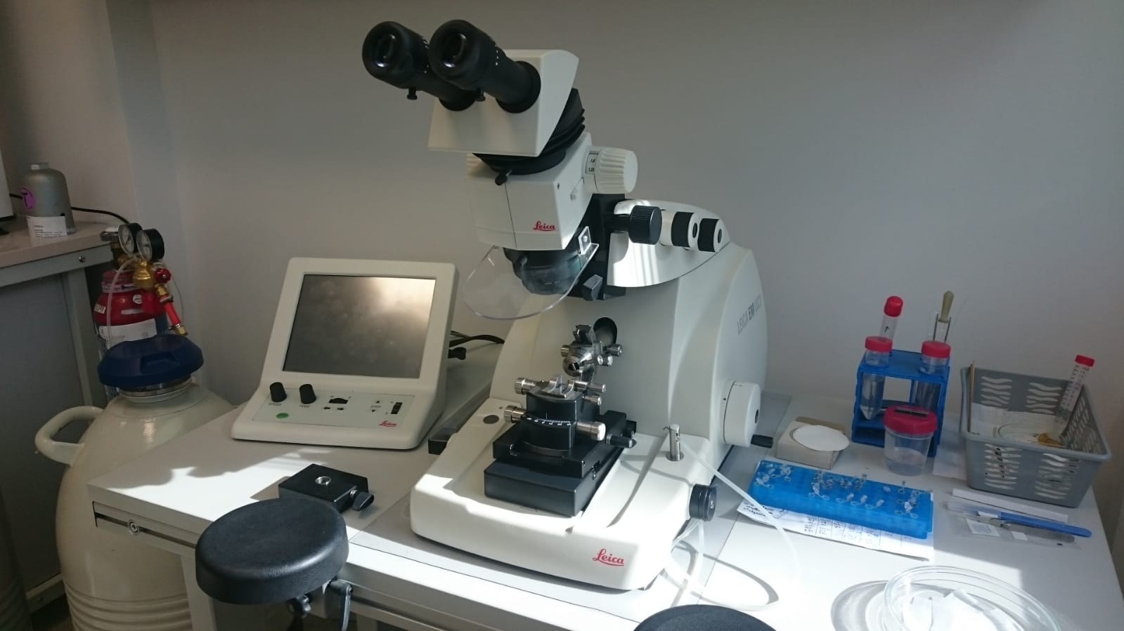
- Leica EM UC7 microtome with FC7 low temperature sectioning syste,
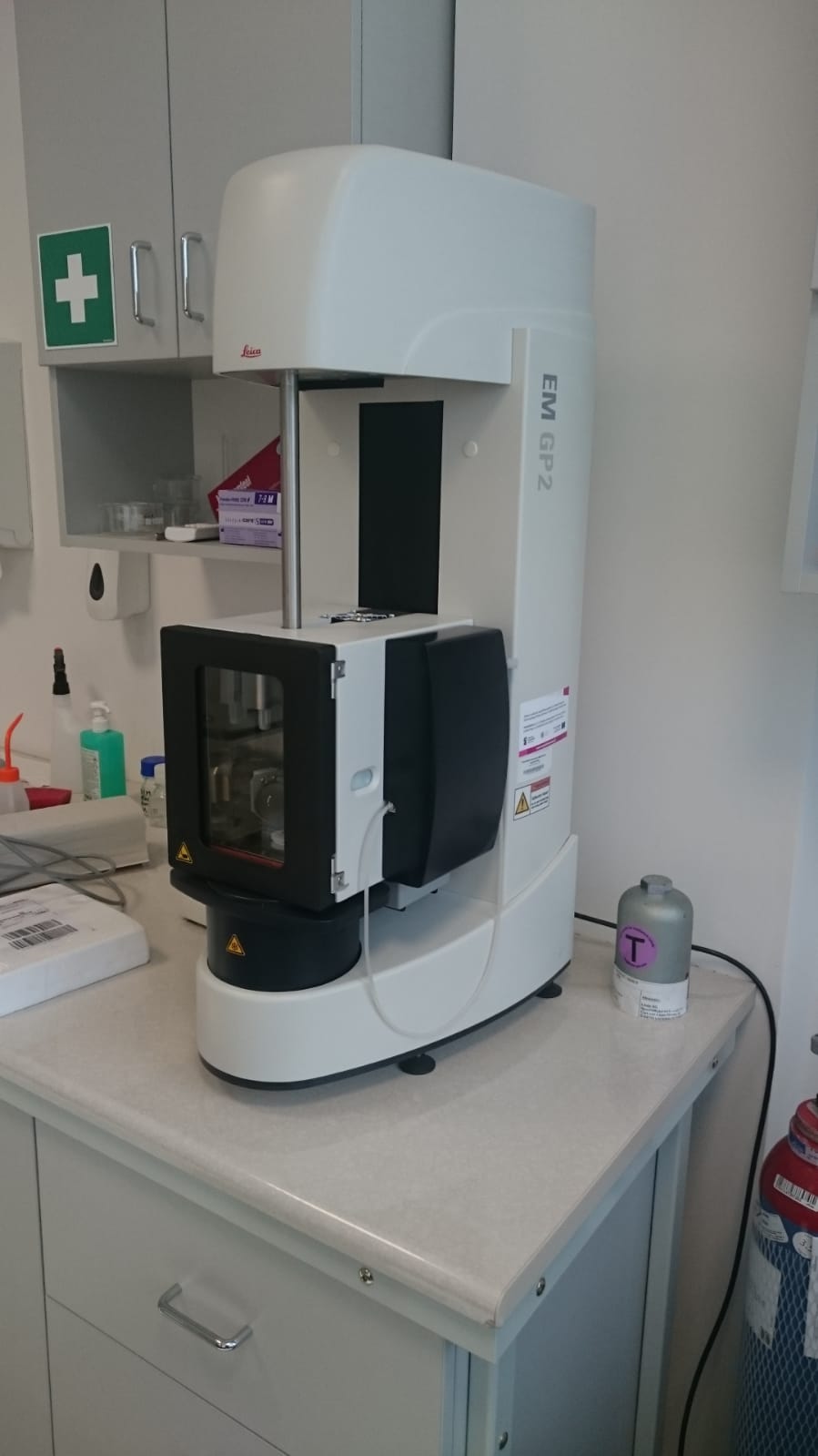
- Leica Automatic Plunge Freezer EM GP2,
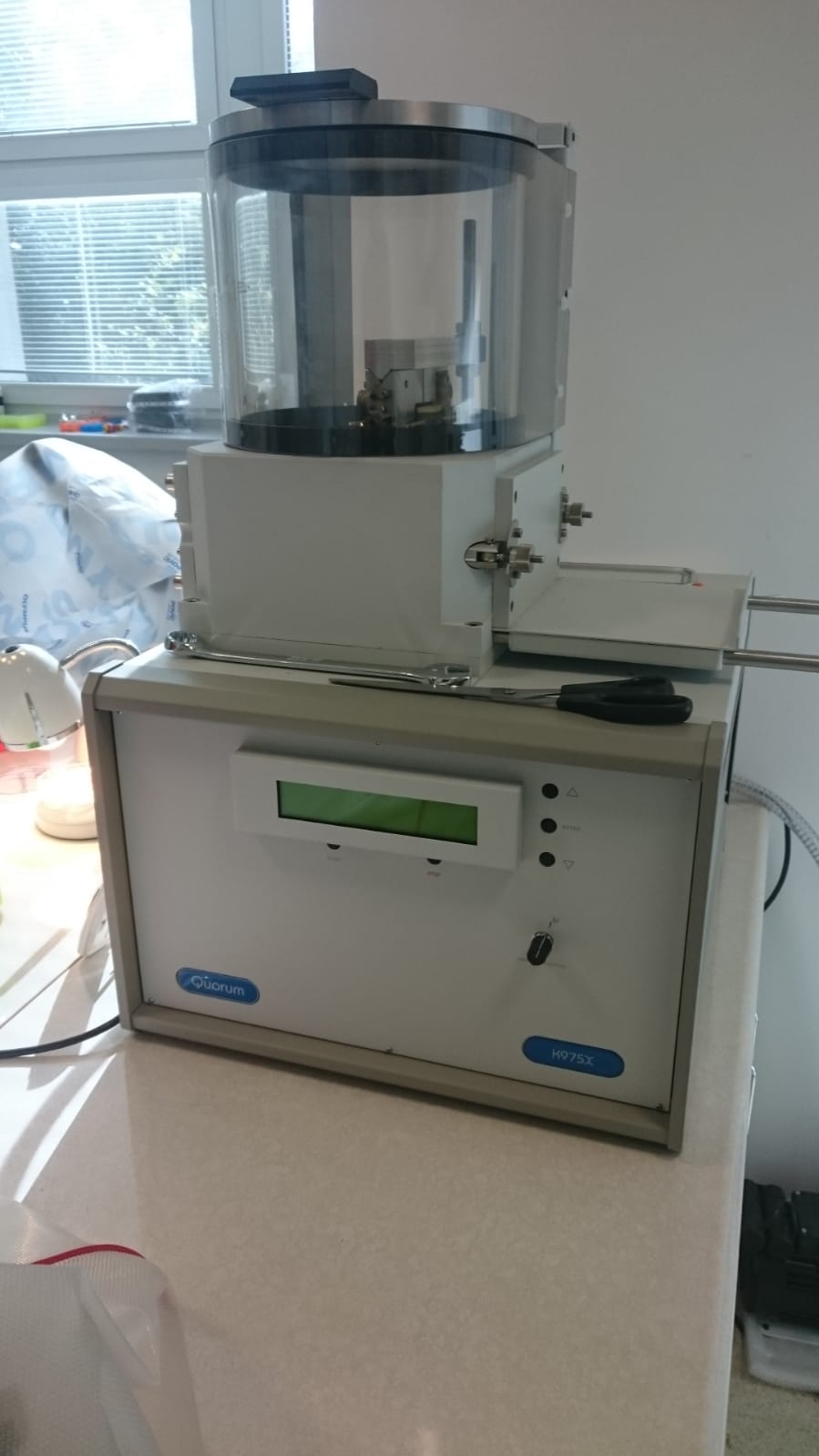
- Quorum Technologies Turbo-Pumped Thermal Evaporator K975X,
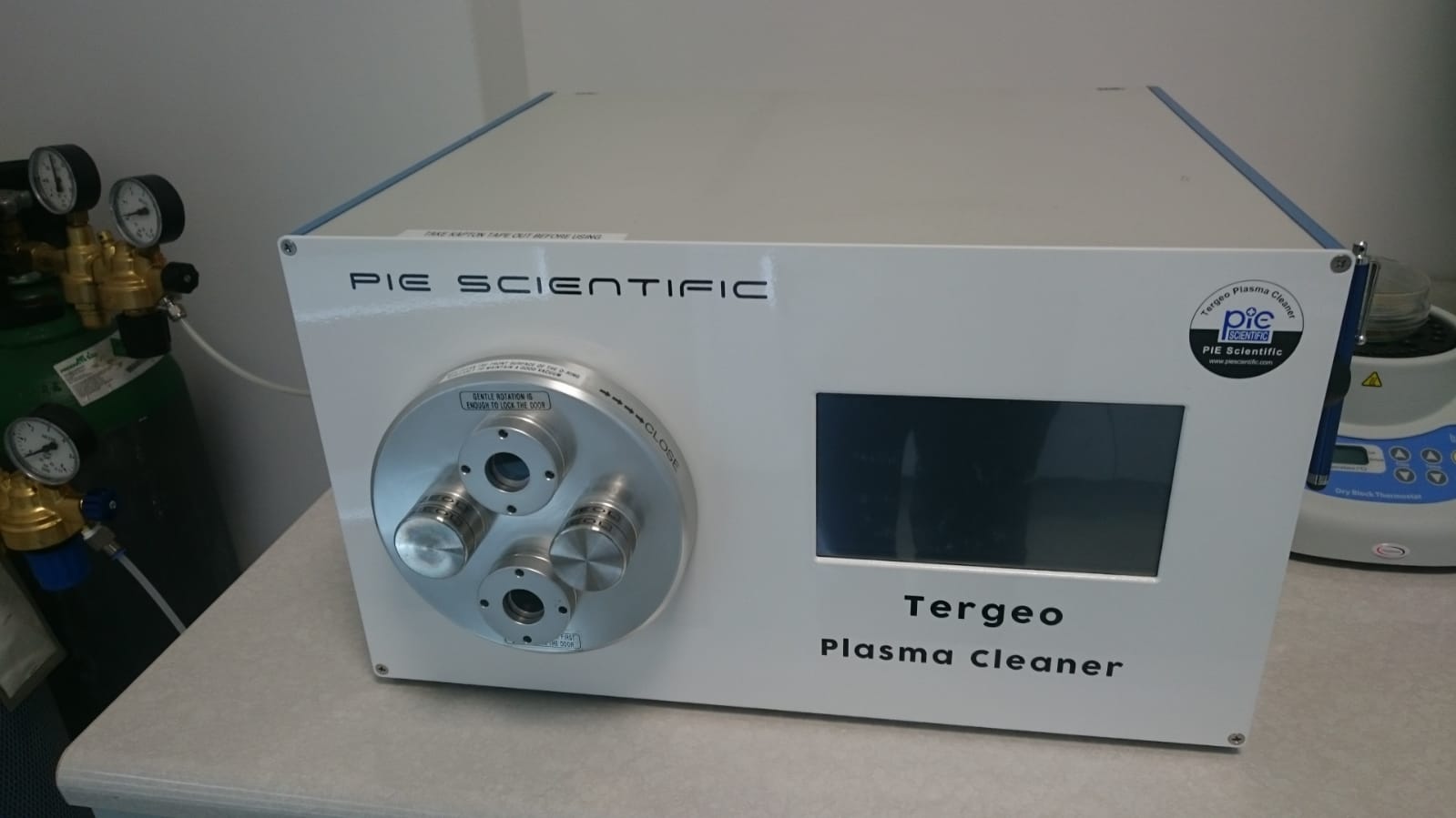
- Plazma cleaner Targeo-EM,
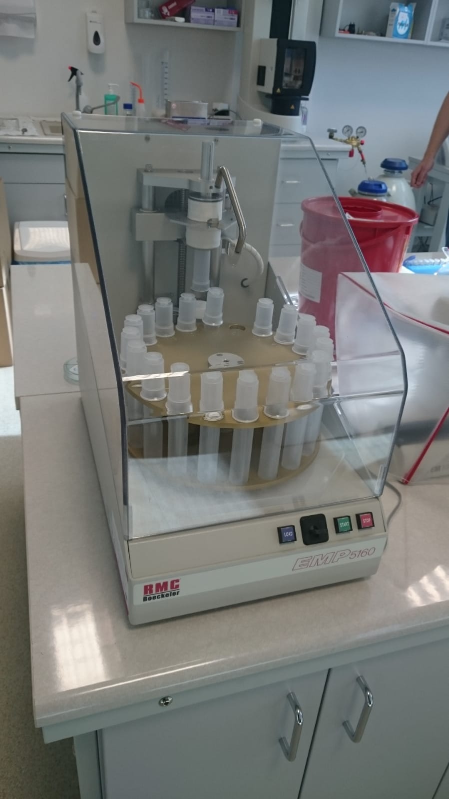
- EMP 5160 Tissue Processor.
Publications
- Markowski A, Migdał P, Zygmunt A, Zaremba-Czogalla M, Gubernator J Evaluation of the in vitro cytotoxic activity of Ursolic Acid PLGA Nanoparticles against Pancreatic Ductal Adenocarcinoma Cell Lines Materials 14 (71), 4917 (2021) https://doi.org/10.3390/ma14174917
Contact
Paweł Migdał Ph. D. pawel.migdal@hirszfeld.pl
Krzysztof Pawlik Ph. D. krzysztof.pawlik@hirszfeld.pl
Gallery